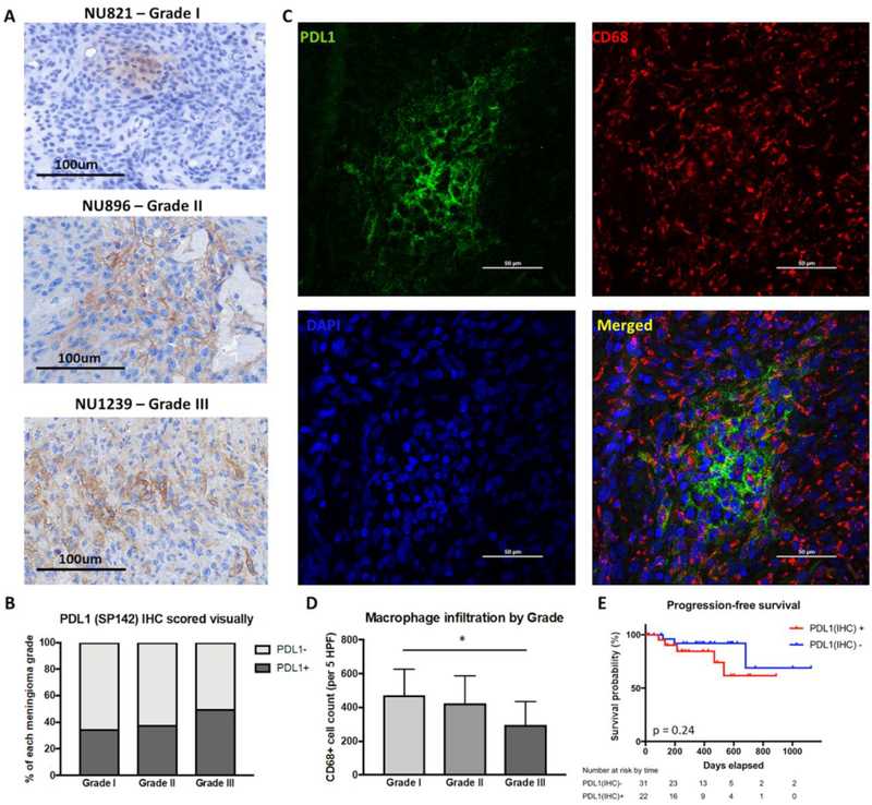
Figure 3: Intratumoral expression of PD-L1 and its impact on outcomes.
(a) Representative IHC staining of PD-L1 in FFPE tumor sections of grade I, II, and III meningiomas. (b) Quantification of intratumoral PD-L1 by grade. Grade III tumors (50%) had greater PD-L1 positivity compared to grade I (35%) and II (38%) tumors. (c) Representative immunofluorescent staining of PD-L1 and CD68 in FFPE tumor sections. (d) Quantification of macrophage infiltration by tumor grade. Compared to grade I meningiomas, grade III tumors had significantly decreased CD68+ cell infiltration (* p < 0.05). (e) Intratumoral PD-L1 was not correlated with PFS.

