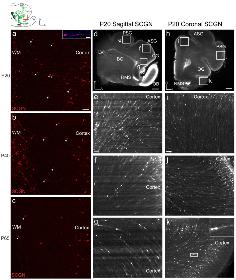
Figure 7. With iDISCO, SCGN+ cells appear to transition between white matter streams and cortex.
a: P20 ferret MMS SCGN+ cells are observed outside the MMS and are oriented towards the prefrontal cortex. b: At P40, fewer SCGN+ cells are observed outside the MMS oriented towards the prefrontal cortex. c: At P65, few SCGN+ cells are observed outside the MMS. d, h: P20 iDISCO cleared sagittal and coronal sections stained with SCGN. e, f, g, i, j, k: Higher resolution images focused on multitude of SCGN+ cells appearing to transition between the white matter streams and cortex. Scale bars a, b, c = 100μm. Scale bar inset a=10μm. Scale bars d, h = 1mm. Scale bars e, f, g, I, j, k=100um.

