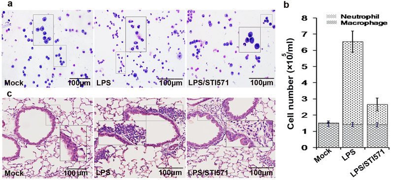
Figure 8. c-Abl activity is responsible for LPS-induced lung inflammation.
(a and b) Visual depiction and quantification of cells in bronchoalveolar lavage fluids. Mice were mock treated or challenged with LPS in the presence or absence of pretreatment with STI571. After 16 h, mice were euthanized, lungs were lavaged and the cell numbers in bronchoalveolar lavage fluids was determined. Ten to sixteen randomly selected fields of view per cytospin slide were photographed (a). Differential cell counts were performed after Modified Wright–Giemsa staining. Data are average ± SD, representative of six experimental animals from one experimental run. (c) STI571 administration blocked LPS-induced sub-epithelium accumulation of leukocytes in lung tissues. Mice were treated as described above. Lung tissue sections were processed for staining with hematoxylin and eosin to examine the sub-epithelium accumulation of leukocytes in lung tissues. Similar results were obtained from at least three independent experiments.

