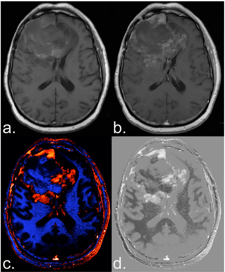
Figure 2.
Benefit of creating a deltaT1 (dT1) map. Shown are the pre (a) and post-contrast (b) T1-weighted images from a patient with recurrent glioblastoma treated with bevacizumab enrolled in the ACRIN 6677 trial. The bright signal on the pre-contrast image and the subtle enhancement on the post-contrast image, makes it difficult to determine the extent of enhancing tumor. Alternatively, the dT1 map created from the difference between calibrated pre- and calibrated post-contrast T1-weighted images clearly delineates enhancing tumor as displayed with either color (c) or in gray scale (d).

