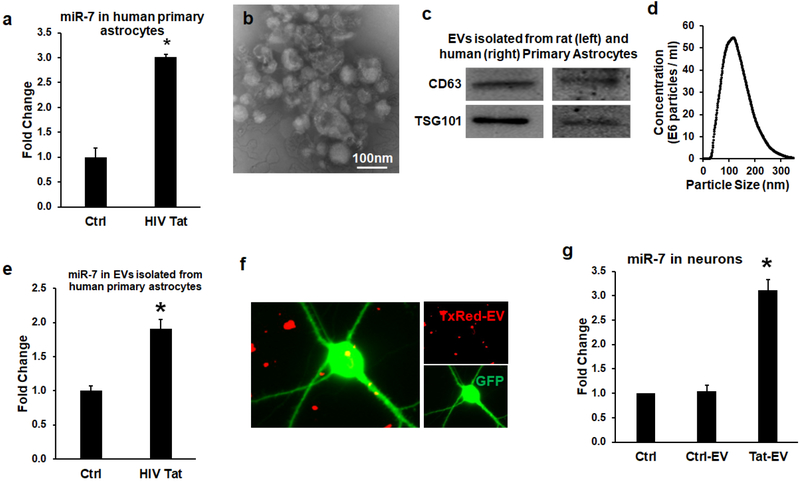
Figure 2. miR-7 was upregulated in the EVs released from HIV-1 Tat-stimulated astrocytes.
(a) Real-time PCR analysis of miR-7 expression in human primary astrocytes stimulated with HIV-1 Tat for 24 h. (b) Western blot characterization of astrocyte EVs. Protein isolated from astrocyte EVs was separated on SDS-PAGE and electroblotted onto nitrocellulose membrane. Blots were probed with exosome marker antibody against TSG101 and CD63. (c) Electron micrograph of EVs isolated from A172 cells. Scale bar=100nm. (d) Size and particle distribution plots of isolated EVs from cell culture by Nanosight Tracking Analysis (NTA). (e) Real-time PCR analysis of miR-7 expression in EVs isolated from HIV-1 Tat stimulated human primary astrocytes for 24 h. (f) Confocal images of rat primary neurons cultured with TxRed labeled ADEVs. Scale bar = 5 μm. (g) Expression levels of miR-7 in untreated (control), EVs from control astrocytes (control-EV), and Tat-stimulated astrocytes (Tat-EV) treated rat primary neurons were measured by qPCR. Bars represent mean ± SD from 3 independent experiments. *p <0.05 vs control..

