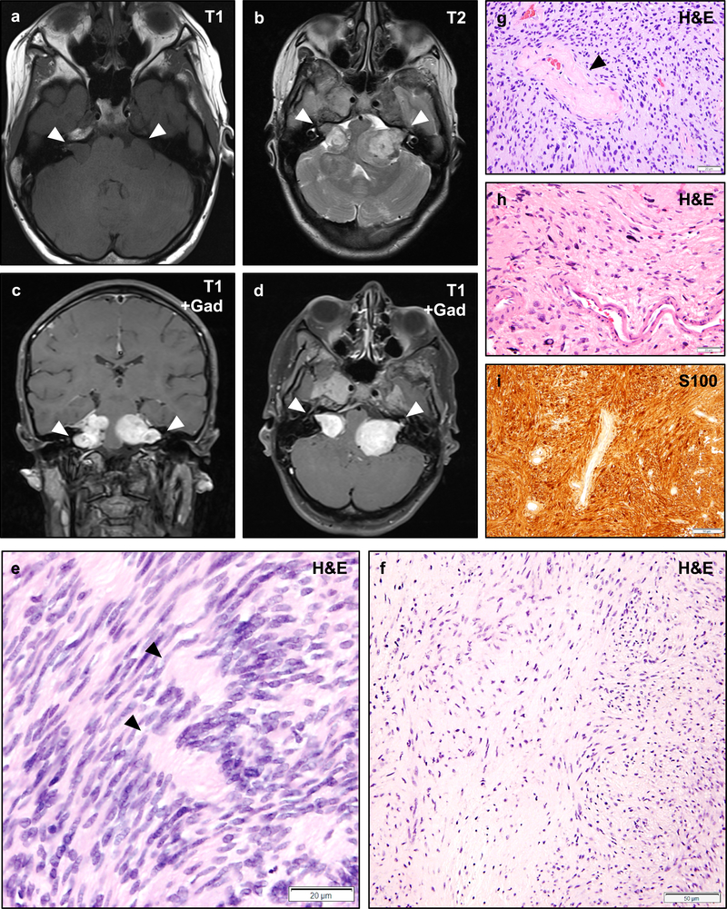
Figure 2 – Vestibular Schwannomas in NF2.
Magnetic resonance imaging (MRI) of an NF2 patient with bilateral vestibular schwannomas involving the cerebellopontine angle and internal acoustic meatus (a–d, arrowheads). Vestibular schwannomas are typically isointense on T1-weighted sequences (a), and show heterogeneous signal on T2-weighted sequences (b). Schwannomas typically exhibit intense gadolinium contrast enhancement (c–d). Histologically, schwannomas consist of spindled cells with tapering ends and eosinophilic to clear cytoplasm which may be architecturally arranged as densely cellular Antoni A regions with focally palisading nuclei that may form alternating layered hypercellular and eosinophilic paucicellular regions known as Verocay bodies (e, arrowheads), or less cellular Antoni B regions with more prominent extracellular matrix which may have a collagenized or myxoid appearance (f). Varying hypercellular and hypocellular regions may correlate with the heterogeneous intensity of schwannomas on T2-weighted sequences (b). Perivascular hyalinization is often a prominent histologic feature in schwannomas (g, arrowhead) and may aid in the diagnosis of lesions with an unusual histologic appearance. Schwannomas frequently exhibit degenerative changes (also known as “ancient change”) including increased pleomorphism, bizarre nuclei, and hyperchromasia (h). Such changes are not known to be associated with an increased risk of recurrence or malignant transformation. Schwannomas exhibit intense and diffuse cytoplasmic and nuclear staining with the S100 antigen (i). Scale bars 20 μm (e, g, h), 50 μm (f, i). A web-interactive tool for viewing images of NF2-related tumors is also available: http://tumoratlas.org/coy-acta-neuropathol-2019.

