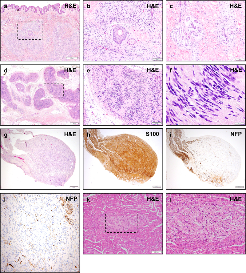
Figure 4 – Histologic Appearance of Non-Vestibular Schwannomas in NF2.
Non-vestibular peripheral schwannomas in NF2 patients may exhibit unusual histologic appearances. Skin biopsy of a cutaneous plaque-like plexiform schwannoma in an NF2 patient shows a nodular and infiltrative proliferation of neoplastic cells involving the dermis and subcutis (a). While sporadic schwannomas are typically circumscribed lesions with a pushing growth pattern, cutaneous schwannomas may infiltrate around skin adnexal structures such as hair follicles and sebaceous glands (b), and deeper eccrine glands and ducts (c). A plexiform schwannoma in the soft tissue of an NF2 patient shows multi-nodular growth along peripheral nerves (d), with typical morphologic features including palisading nuclei and Verocay bodies (e, f). NF2 patients may also develop intraneural schwannomas (g) with whorls of neoplastic cells infiltrating between individual nerve fibers, highlighted by S100 (h) and neurofilament protein (NFP) (i, j) immunohistochemistry. Schwann cell tumorlets may be identified in spinal nerve roots and peripheral nerves, and are hypothesized to represent precursor lesions to larger solitary or plexiform schwannomas (k, l). Scale bars 200 μm (a, d, g, h, i, k), 100 μm (c), 50 μm (b, e, j), 10 μm (f)

