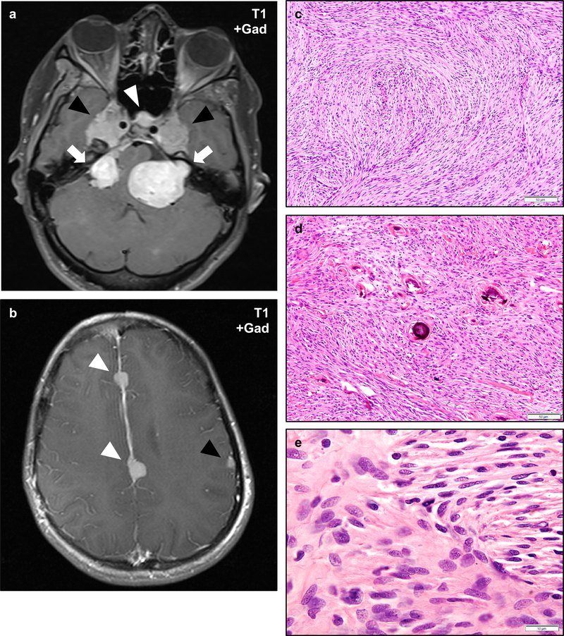
Figure 5 – Meningiomas in NF2.
NF2 patients frequently develop multiple meningiomas, which may involve unusual anatomic locations. Post-gadolinium T1-weight magnetic resonance imaging of the skull base of an NF2 patient with bilateral vestibular schwannomas (a, arrows) and durally based masses consistent with meningiomas in the bilateral Meckel’s caves (black arrowheads) and cavernous sinus (white arrowhead). This patient also had multiple meningiomas involving the falx cerebri (white arrowheads) and cerebral convexity (black arrowhead) (b). Histologically, NF2-associated meningiomas most often exhibit fibrous morphology (c), though any histologic pattern may be encountered. A second NF2-associated meningioma shows scattered psammoma bodies (d). The cells of NF2-associated schwannomas are cytologically similar to sporadic meningiomas and most often exhibit ovoid to spindled cells with find chromatin, scattered nuclear pseudo-inclusions, and mild cytologic atypia (e). Scale bars 50 μm (c, d), 10 μm (e).

