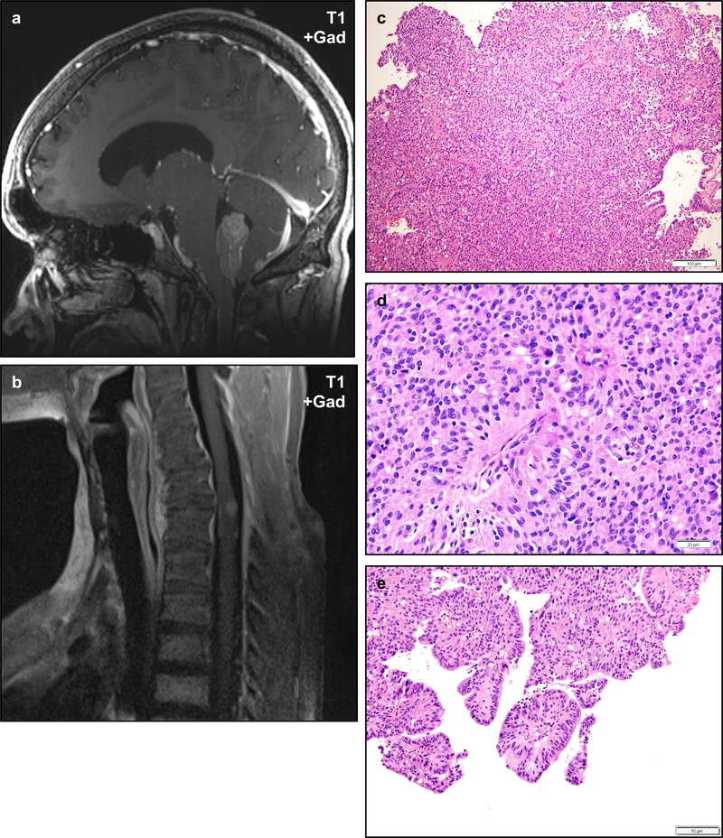
Figure 6 – Ependymomas in NF2.
Ependymomas are frequently diagnosed in NF2 patients and may occur in a variety of locations including unusual sites. Post-gadolinium T1-weighted magnetic resonance imaging of an NF2 patient shows a weakly enhancing intraventricular mass in the fourth ventricle (a). A second NF2 patient developed an intramedullary mass in the cervical spine (b). Histologically, the fourth ventricular mass exhibited typical features of ependymoma including prominent perivascular pseudorosettes and scattered true ependymal rosettes (c, d), with some regions exhibiting prominent papillary architecture (e). Scale bars 100 μm (c), 20 μm (d), 50 μm (e).

