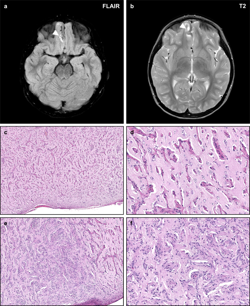
Figure 7 – NF2-associated Meningioangiomatosis.
Meningioangiomatosis is a rare plaque-like leptomeningeal and perivascular proliferation of fibroblastic and meningothelial-appearing cells which may extend along Virchow–Robin spaces. Such lesions are typically hyperintense on FLAIR (a) and T2-weighted sequences (b), with intense contrast enhancement on post-gadolinium T1-weighted sequences. Histologic examination of this lesion from an NF2 patient with refractory epilepsy shows a mixed perivascular proliferation of fibrovascular (c, d) or meningothelial-like cells (e, f) extending from the pial surface into the cortical parenchyma. The intervening cortex shows intact neurons with no definite evidence of focal cortical dysplasia. The neoplastic cells are typically well differentiated with no necrosis, atypia, or prominent nucleoli.

