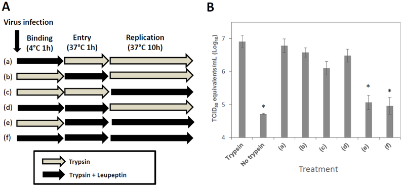
Figure 2.
Addition of leupeptin at different virus replication stages (PEDV KD). Confluent Vero cells were inoculated with PEDV KD at an MOI of 5 in the presence of trypsin (1 μg/ml) or trypsin + inhibitor (leupeptin, 1 μM) at 4C for 1 h (binding stage). After thorough washing with PBS, virus infected cells were transferred to 37C and incubated for 1 h with trypsin or trypsin+inhibitor (entry stage). After another thorough washing, cells were incubated for additional 10 h with trypsin or trypsin+inhibitor before viral replication was assessed. (A) A schematic drawing shows trypsin or trypsin+leupeptin treatment of cells at various stages of virus replication. (B) Virus replication was quantified by real time qRT-PCR at 12h PI. Error bars show standard deviations. Asterisks indicate significant difference (p < 0.05) in virus genome levels, compared to those of PEDV infection with trypsin.

