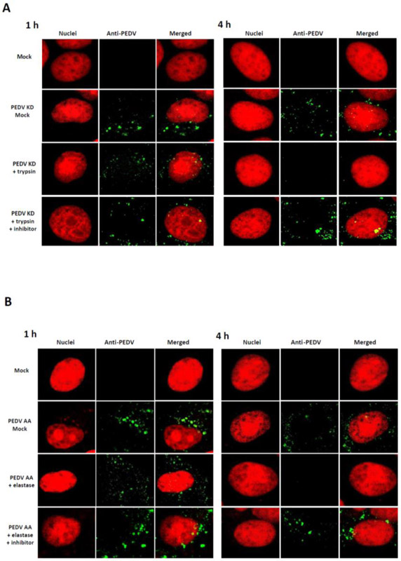
Figure 3.
Confocal microscopy of PEDV entry. Confluent Vero cells grown on Lab-Tek II CC2 chamber slides were infected either with Mock (medium) or PEDV KD (A) or AA (B) at an MOI of 50, and incubated with Mock-medium, trypsin 1 μg/ml (elastase 1 μg/ml) or trypsin+ inhibitor (elastase +inhibitor) for 1 h or 4 h. Fixed cells were probed with swine polyclonal anti-PEDV primary antibodies, followed by FITC-labelled goat-anti-swine antibody (green). Nuclei were stained with sytox orange (5μM) (red), and merged images for PEDV and nuclei were prepared by using Image J.

