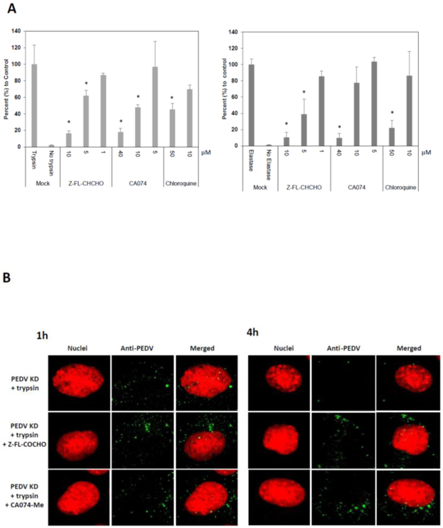
Figure 5.
Effect of Cathepsin inhibitors (Z-FL-CHCHO or CA074-Me) or chloroquine in PEDV entry into the cells. (A). Confluent Vero cells were pre-treated with mock (medium), Z-FL-CHCHO, CA074-Me or chloroquine for 1h before PEDV inoculation (MOI 10). Following virus infection, cells were incubated with same inhibitor in the presence of TPCK-treated trypsin(1 μg/ml) for PEDV KD (left panel) or elastase (1 μg/ml) for PEDV AA (right panel) at 37°C, and total RNAs were collected at 12 h PI. Viral replication was assessed by real time qRT-PCR. (B) Confluent Vero cells grown on Lab-Tek II CC2 chamber slides were pre-treated with mock(medium), Z-FL-CHCHO, CA074-Me, or chloroquine for 1h prior to PEDV inoculation (MOI 50). Then PEDV KD was inoculated to the cells with TPCK-treated trypsin (1 μg/ml). The cells were incubated at 37°C for 1 or 4 h, then fixed and stained for confocal laser scanning microscopy. Error bars show standard deviations. Asterisks indicate significant difference (p < 0.05) compared to the control (trypsin or elastase treatment only).

