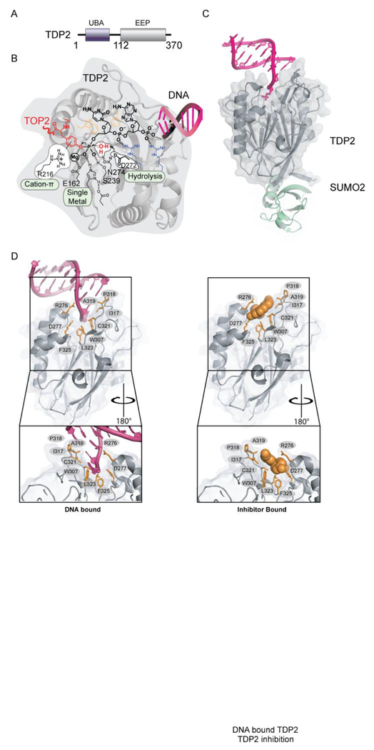
Figure 3: Tyrosyl–DNA Phosphodiesterases-2 (TDP2).
A. Domain organization of TDP2. B. Chemical reactions facilitating direct hydrolysis of a 5’ phosphotyrosyl bond, such as will exist in TOP2-DPC reaction intermediates. C. Mouse TDP2 catalytic domain bound to SUMO PDB ID: 5TVQ. D. Upper left panel, DNA bound TDP2 C-terminal Endonuclease-Exonuclease-Phosphate (EEP) (PDB ID: 5INL) Upper right panel, Inhibitor bound TDP2 EEP (PDB ID: 5J42). Green residues in stick and cartoon show conformation of motif 7 in inhibitor bound state. Residues important for engaging inhibitor or DNA binding are shown in orange sticks. Bottom: 180 degree rotation of upper panels.

