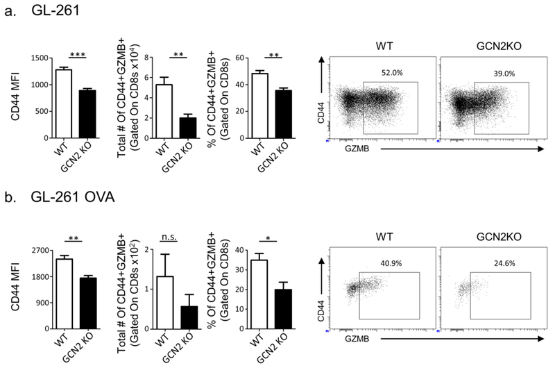
Fig. 2: Glioma infiltrating GCN2 KO CD8+ T-cells are dysfunctional.
GCN2 KO and WT mice were injected with 2x10⁵ GL-261 cells, and after 14 days were euthanized for flow cytometric analysis. In (a) GL-261 injected tumor-infiltrating lymphocytes were stimulated for 5 hours with PMA/Ionomycin, and then stained and analyzed for CD44 mean fluorescence intensity (MFI) and cytokine secretion via flow cytometry. In (b), 1x10⁶ heat shock killed GL-261 OVA cells were injected intraperitoneally into OT-1 and GCN2 KO OT-1 mice. After 14 days of engraftment, CD8+ T-cells from these mice were isolated and transferred i.v. into RAG1 KO mice previously inoculated with GL-261 OVA. Seven days post transfer, mice were sacrificed and analyzed for CD44 MFI and cytokine secretion with flow cytometry. Flow cytometry statistics were calculated, n=5 per group in (a), n=3 per group in (b), representative of two experiments. Unpaired T-test analysis was used to calculate significance. p<0.05*; p<0.01**; p<0.001***. Refer to Supplementary Figure 7b for the gating strategy.

