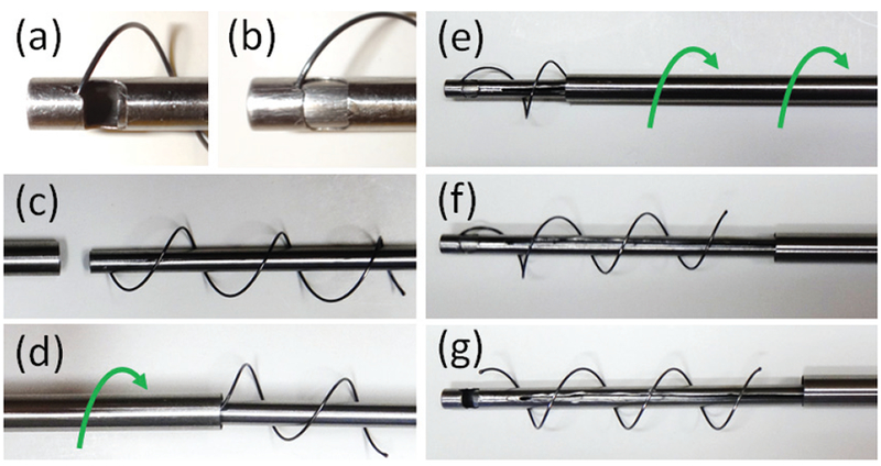
Fig. 10.

Stent delivery tool. (a,b) Prior to delivery, ball on distal end of stent is locked between the inner two cannulas. (c,d) Stent is then compressed and housed inside the outer cannula using a screwing motion. (e) Once distal end of the instrument is placed at desired location in trachea, outer cannula is retracted using an unscrewing motion. (f,g) Retraction of the innermost cannula releases the ball on the distal end of the stent.
