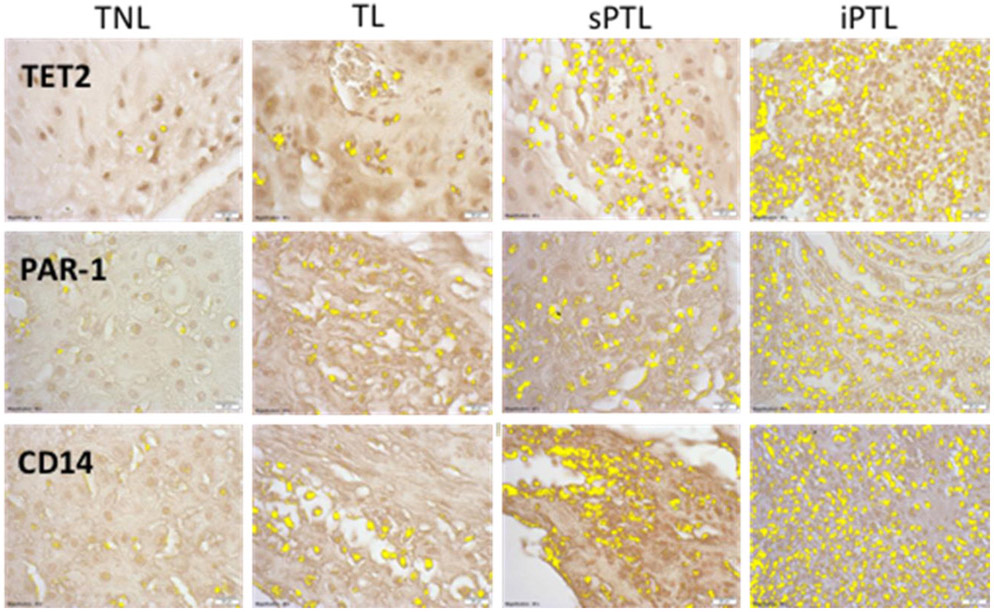
Figure 1.
Representative sections of decidual tissue immunostained for TET2, PAR-1 and CD14 from women who were at term not-in-labor (TNL), women who were at term in labor (TL), women who delivered spontaneously preterm with no clinical signs of infection (sPTL) and women who delivered preterm with PPROM and clinical signs of infection (iPTL). Specific antigen staining was highlighted in yellow using the Measuring Images tool in the cellSens software. There was little staining for TET2, PAR-1 or CD14 in women with TNL. Staining increased slightly for TL but was markedly increased in sPTL and iPTL. The increasing pattern of staining for TET2 and PAR-1 correlated with the macrophage marker, CD14. Decidual cells did not expression TET2 or PAR-1. All pictures and analyses were done with a 40X lens.

