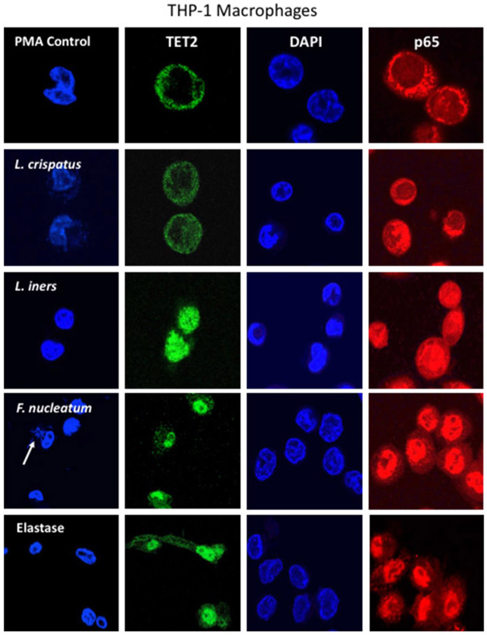
Figure 7.
Representative images of the nuclear translocation of TET2 (green) and the p65 subunit of NF-κB (red) in THP-1 macrophage cells in response to bacteria (100:1, bacteria to cells) or elastase (0.033 U/ml). DAPI blue indicates the location of the nucleus. In control cells, TET2 and p65 were primarily localized to the cytosol indicating TET2 and NF-κB were not activated in most cells. In cells treated with L. crispatus, there was some movement of TET2 and p65 from the cytosol to the nucleus, however in cells treated with L. iners, F. nucleatum or elastase, TET2 and p65 were primarily localized to the nucleus in most cells. White arrow for F. nucleatum points to nuclei of bacteria engulfed by a THP-1 macrophage.

