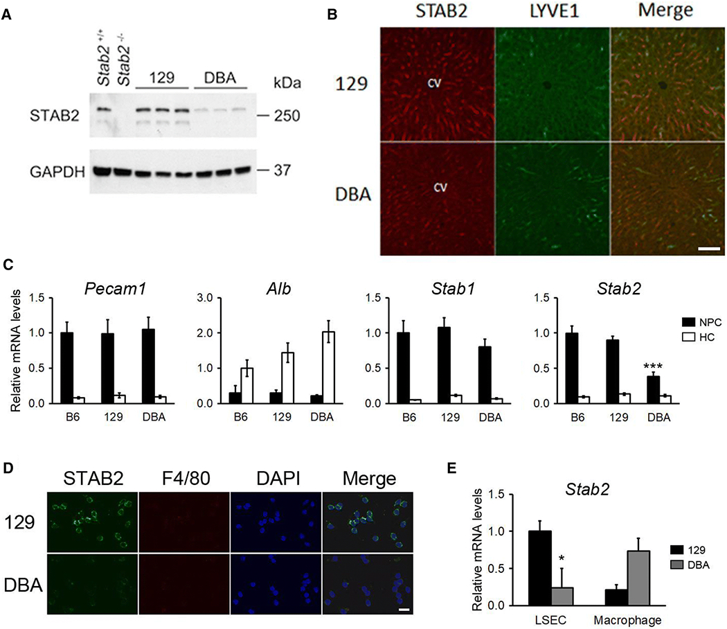
Fig 7. Reduced expression of Stab2DBA in the liver sinusoidal endothelial cells (LSECs) of DBA/2J.

(A) A western blot for STAB2 from the whole liver of 129S6 and DBA/2J mice. Liver lysates from Stab2+/+ and Stab2−/− mice on a C57BL/6J background were used as a positive and negative control. (B) Immunohistochemical staining of STAB2 in the liver from 129S6 and DBA/2J mice. STAB2 signal was detected along sinusoids and partly overlapped with Lymphatic vessel endothelial hyaluronan receptor 1 (LYVE1). Bar = 100 μm. (C) Quantitative RT-PCR of Pecam1, Albumin (Alb), Stab1 and Stab2 in the non-parenchymal cells (NPCs) and hepatocytes (HCs) isolated form the liver of 3 months old C57BL/6J, 129S6 and DBA/2J male mice (n = 5 each. ***p < 0.001 vs. C57BL/6J and vs. 129S6). (D) Immunofluorescence staining for STAB2 and a macrophage marker F4/80 of LSECs isolated from the liver of 129S6 and DBA/2J mice. Nuclei were visualized by DAPI staining. Bar = 20 μm. (E) Quantitative RT-PCR of Stab2 in the isolated LSEC and macrophage fractions from the livers of 129S6 and DBA/2J. *p < 0.05 vs. 129S6 in LSEC (n = 3). Macrophage expression was tended to be higher in DBA, but statistics were not performed because sample size in DBA2J was n = 2 while n = 3 in 129S6.
