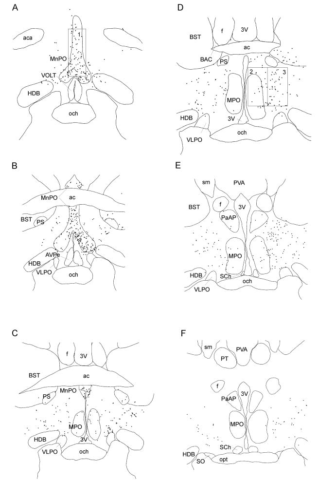
Figure 3.
Drawings of brain sections from rostral to caudal levels of preoptic area after CTb injection into the RMR. Each dot represents one retrogradely-labeled neuron. The counting box for the MnPO was placed dorsal to the opening of the 3rd ventricle just rostral to the anterior commissure (A, box 1). Neurons in the medial preoptic area were counted by placing a box with the ventral border 300 μm above the surface of the optic chiasm and its medial border along the wall of the 3rd ventricle (D, box 2) at the level of the bed nucleus of the anterior commissure of Gurdjian (BAC). For the DLPO, the counting box was placed just lateral to the medial preoptic box (D, box 3). 3V: third ventricle; ac: anterior commissure; aca: anterior commissure, anteriot part; AVPe: anteroventral periventricular nucleus; BST: bed nucleus of the stria teminalis; f: fornix; HDB: nucleus of the horizontal limb of the diagonal band; MnPO: median preoptic nucleus; MPO: medial preoptic nucleus; och: optic chiasm; opt: optic tract; PaAP: paraventricular hypothalamic nucleus, anterior parvicellular part; PS: parastrial nucleus; PT: paratenial thalamic nucleus: PVA: paraventricular thalamic, anterior part; SCh: suprachiasmatic nucleus; sm: stria medullaris of the thalamus; SO: supraoptic nucleus; VLPO: ventrolateral preoptic nucleus; VOLT: vascular organ of the lamina terminalis.

