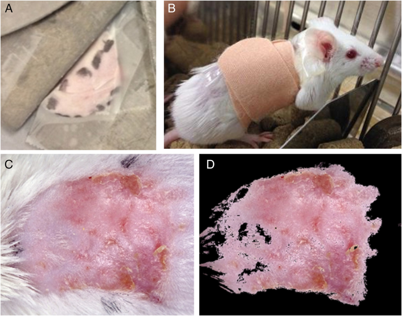
Figure 1.
Radiation exposure, treatment delivery, and quantification of treatment response. (A) The dorsal skin of the mice was pulled through a slit in the lead shielding and taped to the surface of the lead to create a 2 cm diameter localized cutaneous radiation injury (CRI). The irradiated area was outlined with a marker so that the treatment could be applied to the irradiated area prior to the appearance of radiation dermatitis. (B) After application of 0.3 ml of either the treatment or vehicle, the irradiated area was covered with a bandage. (C) Image of a typical CRI at 4 weeks following radiation exposure and treatment with vehicle alone. (D) Same image as shown in (C) following automatic segmentation to quantify wound area.

