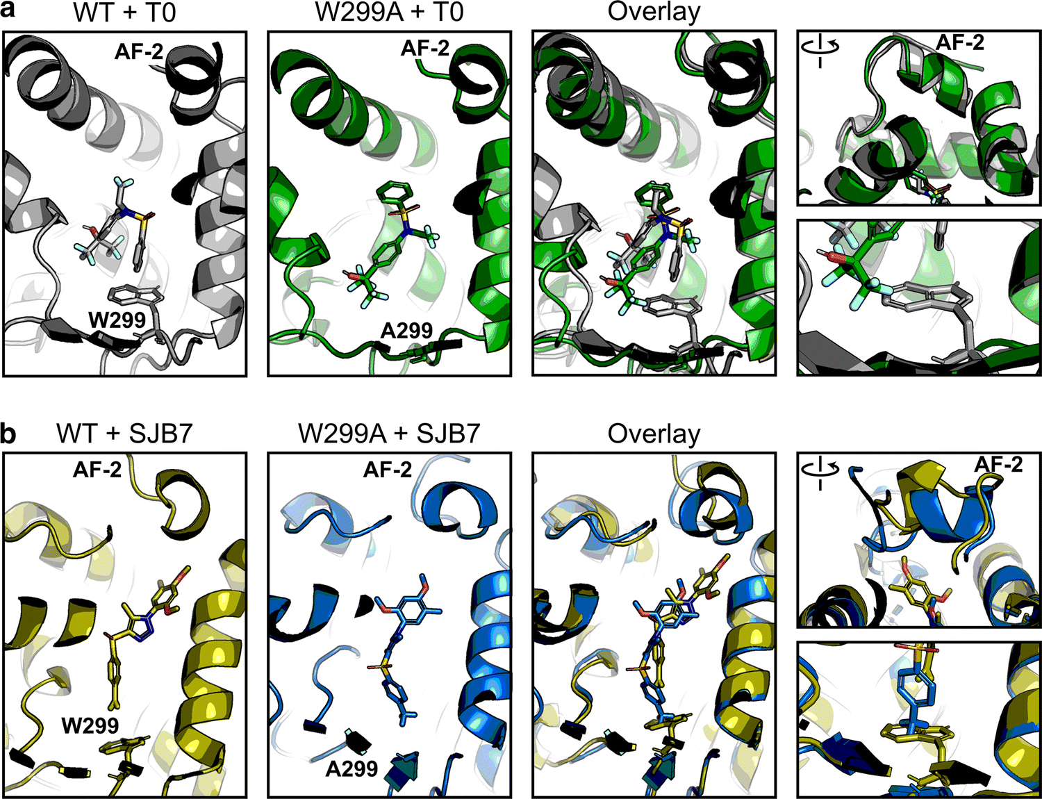
Fig 4. The W299A mutation does not affect agonist-bound PXR conformation.

Molecular dynamics simulations were performed for WT and W299A PXR LBD in the presence of (a) T0 or (b) SJB7. Individual and overlaid images are shown for the ligands in the ligand-binding pocket; the right panels focus on the AF-2 helix and W/A299. Each panel is derived from the same frame of its respective simulation. Protein-ligand structures are shown in gray (WT + T0), green (W299A + T0), olive (WT + SJB7), and blue (W299A + SJB7). Ligand heteroatoms are shown in red for oxygen, blue for nitrogen, yellow for sulfur, and teal for fluorine
