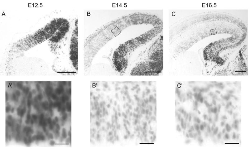
Figure 1. Beta-catenin signaling levels undergo progressive reduction during cortical neurogenesis.

Comparable coronal sections from E12.5, E14.5, and E16.5 BAT-gal mouse cortex after X-gal staining reaction. BAT-gal mice express β-galactosidase in response to β-catenin signaling. Additional detail of the ventricular zone is shown in high power insets below. Scale bars are 100 μm in low power images and 20 μm in high power images.
