Figure 3.
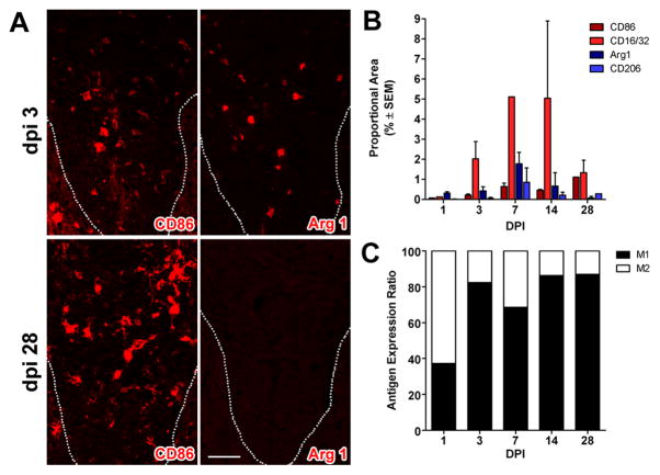
M1 macrophages dominate zones of Wallerian degeneration after SCI. (A) Similar to the macrophage response that evolves at the lesion epicenter (see Fig. 2), M1 (CD86+) and M2 (Arg-I) macrophages co-exist within the dorsal funiculus (DF) at 3 dpi but only M1 macrophages persist until 28 dpi (dotted line indicates the border of the gray matter and dorsal funiculus). (B) Quantitation of macrophages expressing M1 and M2 markers in the DF at different times post-SCI. (C) When expressed as a ratio of M1:M2 cells, there is an obvious shift towards a M1 macrophage by 3 dpi. (M1 and M2 markers; red, AF546 and nuclear stain with DAPI; blue); scale bar = 20μm.

