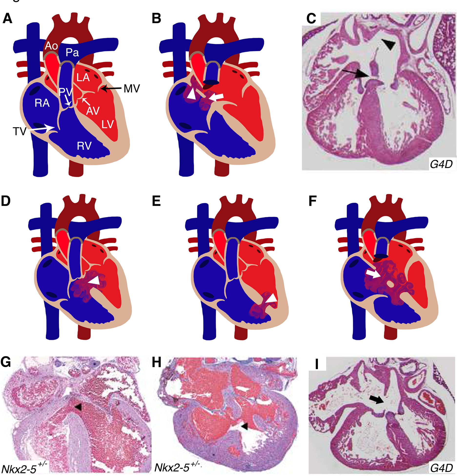
Figure 1. Murine models of cardiac septation defects:

(A) Illustration depicts a cross-sectional view of a normal and mature four chambered heart. (B) Location of primum (arrowhead) and secundum (arrow) ASD in human heart diagram which has been recapitulated in Gata4Δex2/WT (G4D) mouse model (C). (D, E) Schematics display similar features of perimembranous and muscular VSDs (arrowhead) seen in human CHD patients were also detected in Nkx2–5+/− mice (G, H). (F) The diagram represents AVSD phenotype with single valve (arrow) noted in humans, resembled by the G4D murine model (I). RA: right atrium; RV: right ventricle; LA: left atrium; LV: left ventricle; MV: mitral valve; AV: aortic valve; PV: pulmonary valve; TV: tricuspid valve; Ao: Aorta; Pa: Pulmonary artery; ASD: atrial septal defect; VSD: ventricular septal defect; AVSD: atrioventricular septal defect. (C, F, I) and (G, H) have been adapted from Rajagopal et al. 2007 and Winston et al. 2010 with permission.
