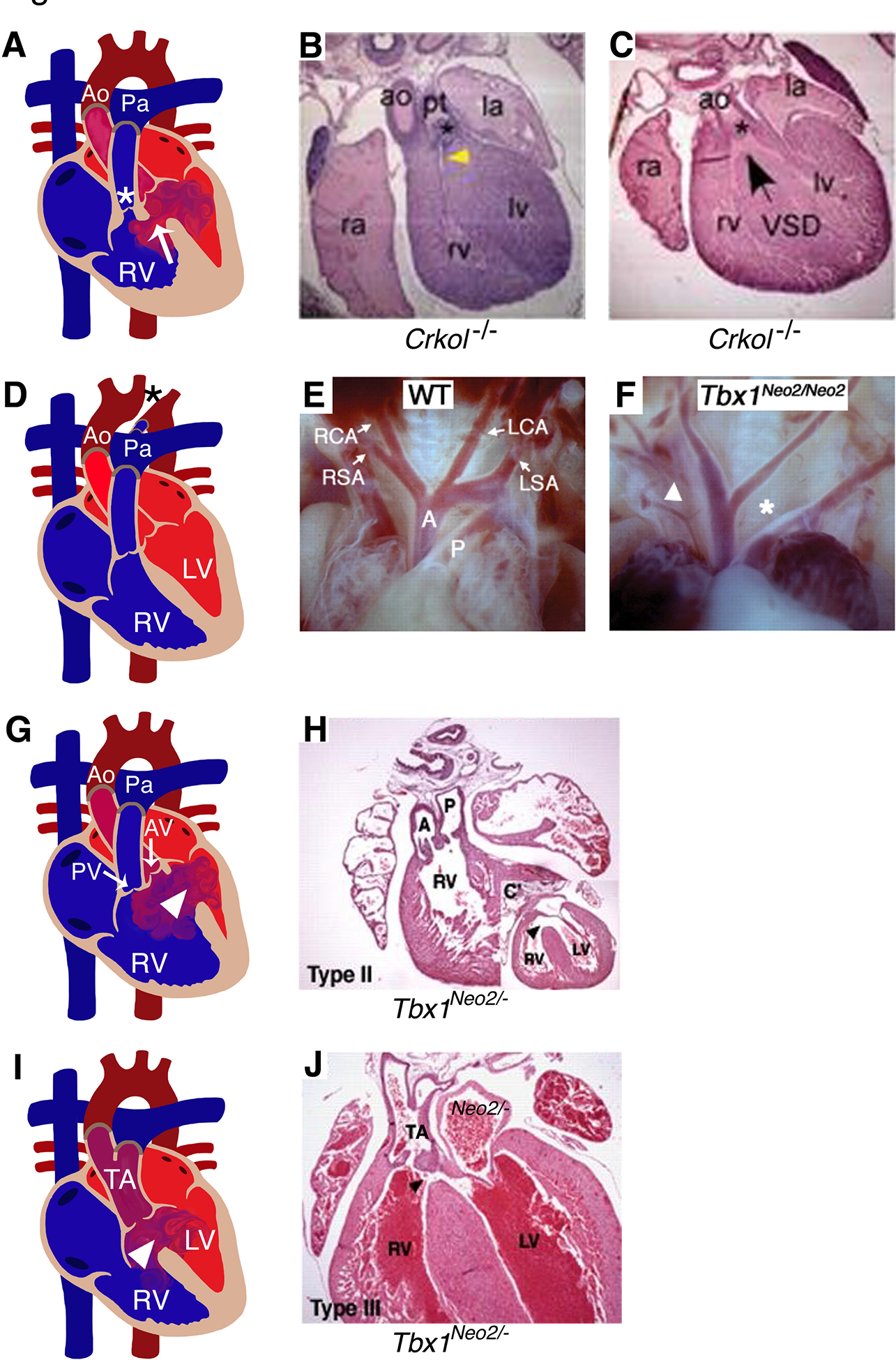
Figure 3. Murine models for conotruncal and aortic arch artery defects.

(A) Diagram illustrates TOF, characterized by right ventricular hypertrophy, VSD (arrow), stenosis of pulmonary valve and sub-valvar area (*) in human heart. Comparable TOF phenotype was observed in mouse model of Crkol−/− at embryonic day 16.5 shown in B and C. (D, F) IAA (*) detected in human patients were recapitulated in Tbx1Neo2/Neo2 mouse model at E18.5 compared to wildtype mouse (E). (G, I) Representative diagram of a diseased heart display DORV and TA with VSD (arrow head) in human (G, I). Similar phenotypes were observed in Tbx1Neo2/Neo2 mice (H, J) at E18.5. RA: right atrium; RV: right ventricle; LA: left atrium; LV: left ventricle; AV: aortic valve; Ao/A: Aorta; Pa/P: Pulmonary artery; TOF: tetralogy of Fallot; IAA: Interrupted aortic arch; DORV: double outlet right ventricle; TA: truncus arteriosus; VSD: ventricular septal defect. (E, F, H, J) have been adapted from Guris et al. 2001 and Zhang and Baldini 2008 with permission.
