
Figure 1.
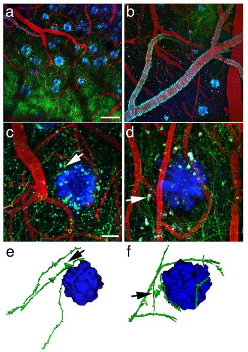
Amyloid plaques alter the morphology and trajectory of neurites in vivo. Low magnification images (a and b) provide an overview of the GFP-AAV injection site containing GFP-filled neurites (green), blood vessels containing Texas red, and amyloid deposition stained with methoxy-XO4 (blue). The angiogram, plaques, and cerebral amyloid angiopathy (seen coating a large vessel in b) are easily identified from week to week allowing re-imaging of the same sites over different imaging sessions. At higher magnification (c and d), we observe dystrophic neurites (arrows) associated with plaques. We also confirm that dendrites do not penetrate plaques, rather their trajectory curves around plaque edges. Three-dimensional reconstructions of plaques and neurites (e and f) clearly show this curvature around plaques and highlight dystrophies near plaques (arrows). Scale bars a and b 100 μm, c and d 20 μm.