
Figure 5.
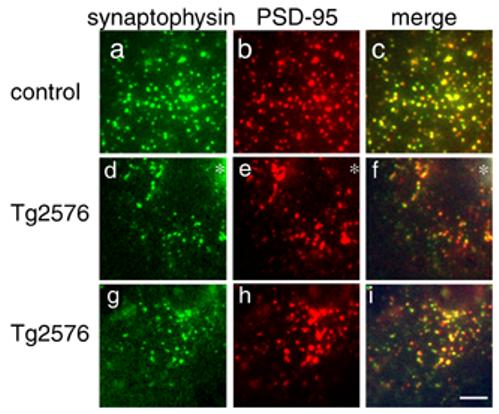
Spine loss reflects loss of excitatory synapses. Immunostaining for synaptophysin (a, d, g) and PSD-95 (b, e, h) reveals similar levels of co-localization (c, f, i) of these pre and postsynaptic markers in control cortex, Tg2576 cortex near the edge of dense plaques (asterisks in d, e, f), and in Tg2576 cortex distant from plaques (g, h, i). Similar amounts of co-localization indicate that dendritic spine loss in Tg2576 cortex is accompanied by loss of the presynaptic element, and not retraction of spines leaving a solitary presynaptic element. There are also qualitatively fewer synapses in Tg2576 cortex near plaques than in control, implying a loss of excitatory synapses concomitant with spine loss. Scale bar 5 μm.