
Figure 6: Photolytic lesions in mmp-9 mutants results in the over production of injury-induced progenitors and regenerated photoreceptors.
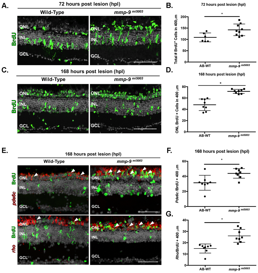
(A) BrdU-labeled cells (green) in wild-type and mmp-9 mi5003. (B) Number of BrdU+ cells from wild-type (109 ± 19.66 cells; n=6) mutant retinas (142.3 ± 25.72 cells; n=9) at 72 hpl. *p = 0.0186. (C) BrdU-labeled cells (green) in wild-type and mutant retinas at 168 hpl. (D) Number of BrdU+ cells in the ONL of wild-type (48 ± 10.24 cells; n=8) and mutant retinas (71.71 ± 3.7 cells; n=8). *p=0.0001. (E) Double labeled, regenerated photoreceptors using in situ hybridization for rods (rho) and cones (pde6c; red signal) and BrdU (green) at 168hpl. (F) Number of regenerated cone photoreceptors in wild-type (31.42 ± 9.88 cell; n=8) and mutant retinas (43.83 ± 10.68 cells; n=8) at 168 hpl. *p=0.0301. (G) Number of regenerated rod photoreceptors in wild-type (14.88 ± 4.02 cells; n=8) and mutant retinas (26.04 ± 5.69 cells; n=8) at 168 hpl. *p=0.0005. ONL- outer nuclear layer; INL- inner nuclear layer; GCL- ganglion cell layer. Scale bars equal 50μm.