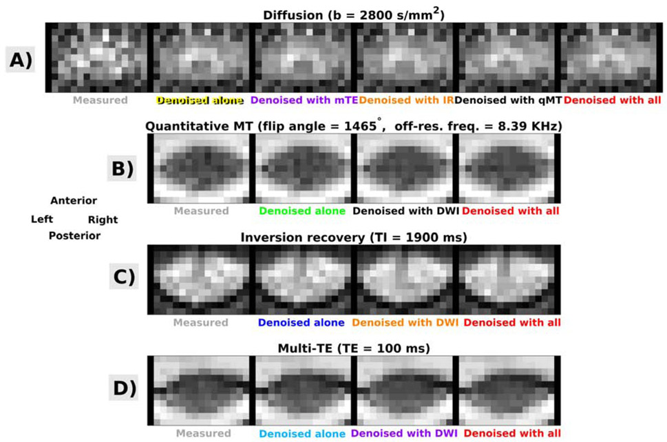
Fig. 3.
Examples of MP-PCA denoising in one subjects who was scanned with vendor 1. Panels A, B, C, D show raw and denoised images, obtained according to different strategies. DW imaging: panel A; qMT imaging: panel B ; IR imaging: panelC; mTE imaging: panelD. Anterior, Posterior, Right, Left respectively indicate subject’s anterior, posterior parts and right and left sides.

