
Figure 4. Cells containing μdot [PIN+] show transient large aggregates.
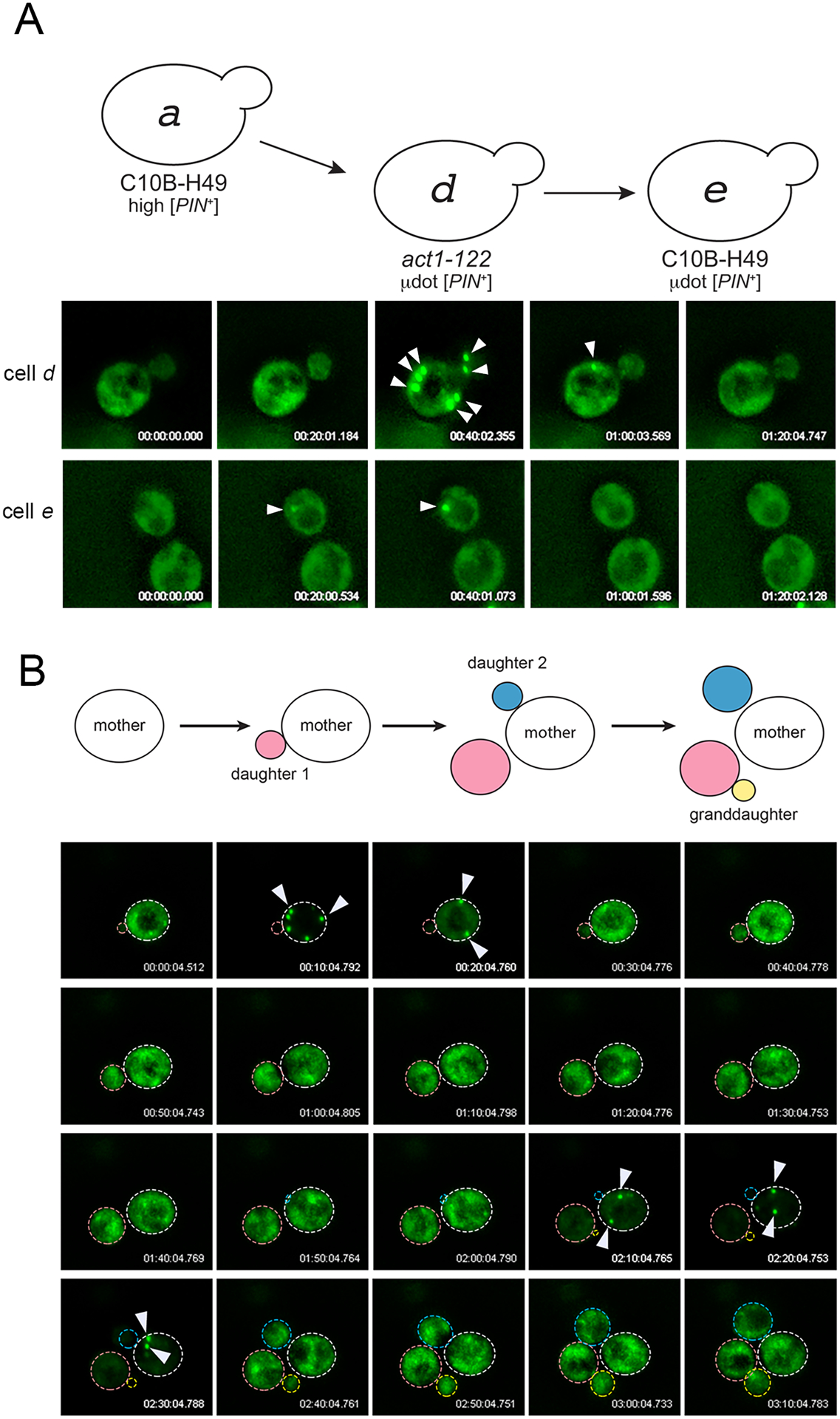
A. 3D-time-lapse imaging of cell d (act1–122) or cell e (wildtype C10B-H49) containing μdot [PIN+]. The appearance and disappearance of large aggregates are indicated with arrowheads. B. A 3D time-lapse of a single dividing cell shows the appearance and disappearance of large aggregates over several cell divisions. All 3D time-lapse imaging was done without addition of copper sulfate, such that low Rnq1-GFP fluorescence reflects trace amounts of copper sulfate in the synthetic complete media (see Experimental Procedures).