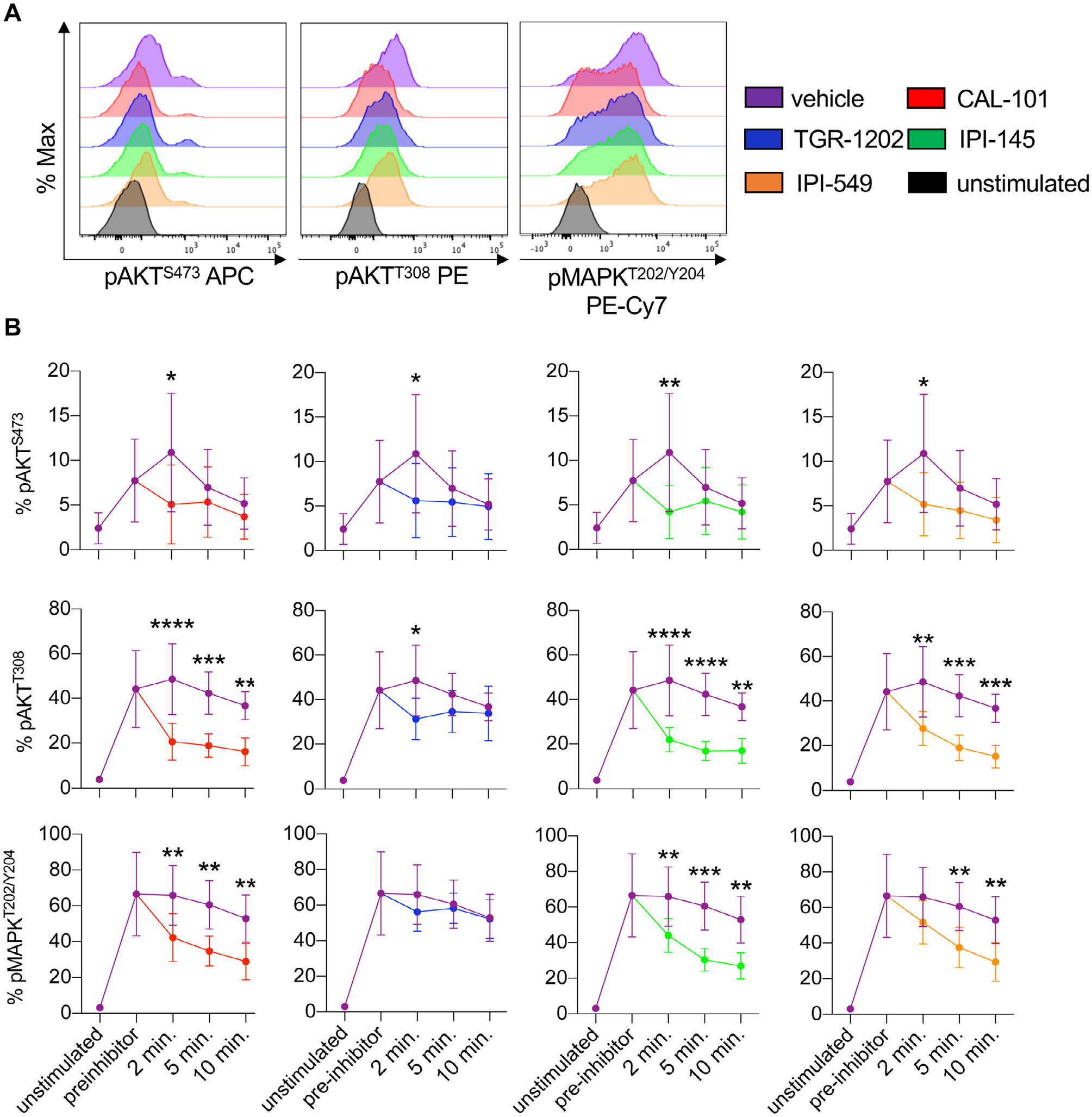
Figure 2.

PI3Kγ and/or PI3Kδ inhibitors target the PI3K/AKT/MAPK signaling axis in CD8+ T cells. Pmel-1 splenocytes were stimulated with 1 μM hgp100 for 3 h and then treated with 10 μM PI3K inhibitors. Phosphorylation was analyzed at 2, 5, and 10 min post inhibitor addition. (A) Representative flow cytometry plots of CD8+ T cell phosphorylation of AKTS473, AKTT308, and MAPKT202/Y204 2 min post inhibitor addition. (B) Phosphorylation kinetics of these residues before and after inhibitor addition comparing vehicle to PI3K inhibitor-treated cells. Data analyzed by unpaired two-tail t-tests at each time point, n = 6 mice/group from two independent studies. All data are represented as the mean ± the SD with statistical significance as p < 0.05*, p < 0.01**, p < 0.001***, and p < 0.0001****.
