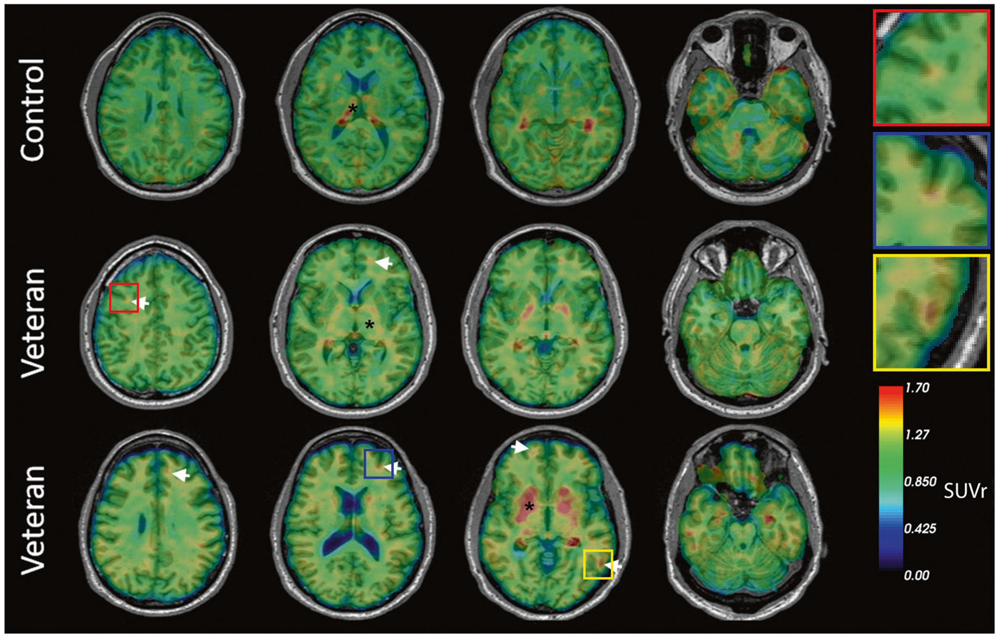
Fig. 3. Representative transaxial brain images of [18F]AV1451 PET of veterans with history of multiple blast exposures.

Veterans 1 and 2 show cortical ligand retention at the white/gray matter junction, as is characteristic of the distribution of tauopathy in CTE (white arrows). * Indicates areas of non-specific binding and uptake. The top row represents a cognitively healthy control. Insets show higher magnification of foci of [18F]AV1451 ligand retention.
