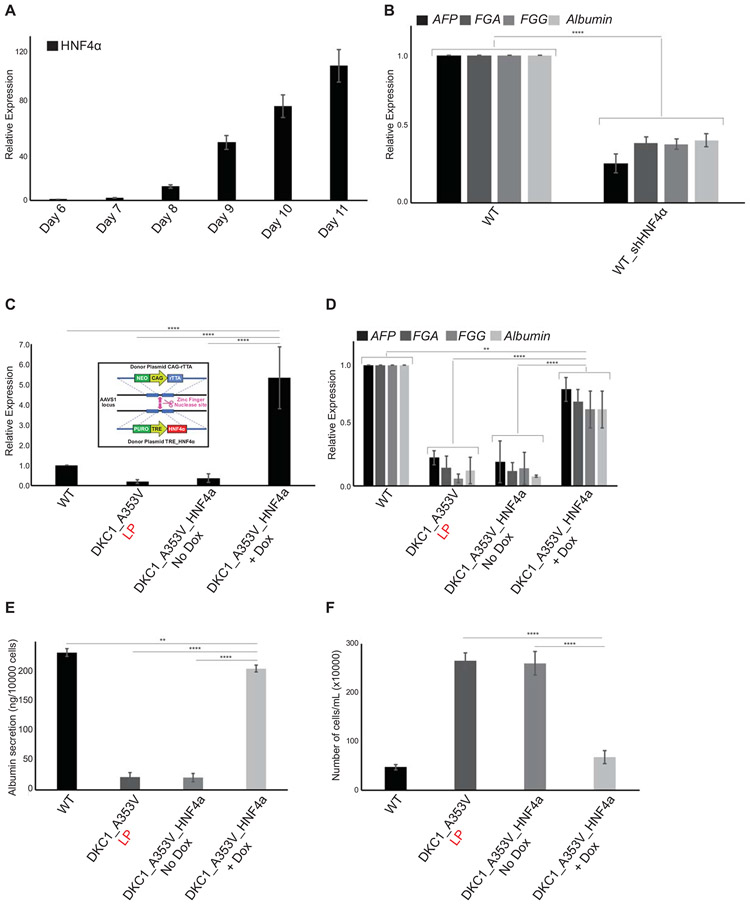
Figure 5: HNF4a expression rescues hepatocyte development in DKC1_A353V mutant cells that retain short telomeres.
(A) HNF4α expression during hepatocyte derivation from WT hESCs. HNF4α expression increases until Day 11 of differentiation (hepatic endoderm stage). (B) Relative expression of hepatocyte markers by real-time quantitative PCR after 21 days of differentiation (mature hepatocyte stage) in WT and WT_sh HNF4α cells. (C) HNF4α expression in WT, DKC1_A353V_LP, and conditional DKC1_A353V_ HNF4α cells with or without addition of Doxycycline. Inlet: construction and cloning of conditional HNF4α cassette into the AAVS1 locus of DKC1_A353V hESCs. (D) Relative expression (by real-time quantitative PCR) of hepatocyte markers after 21 days of differentiation in WT, DKC1_A353V_LP, and conditional DKC1_A353V_ HNF4α cells with or without addition of Doxycycline. (E) Quantification of albumin secretion after 21 days of differentiation in WT, DKC1_A353V_LP, and conditional DKC1_A353V_ HNF4α cells with or without addition of Doxycycline. (F) Total number of cells after hepatic differentiation of WT, DKC1_A353V_LP, and conditional DKC1_A353V_ HNF4α cells with or without addition of Doxycycline. Cells were collected on Day 21 and figure shows total number of cells found in each population (total numbers quantified by cell counter). n=3, mean ± SEM, *p≤0.05; **p≤0.0025; ***p≤0.001; ****p≤0.0001. Statistical analysis was performed using one-way ANOVA followed by Tukey’s post hoc test.

