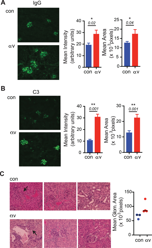
Figure 5: αv-deletion increases kidney antibody deposition and glomerulonephritis:
(A-B) Glomerular immune complex deposits determined by immunofluorescence staining for IgG (A) and complement C3 (B). Left panels show representative images from 7–8 week old control Tlr7.1 tg and αv-CD19.Tlr7.1 tg mice. Right panels show intensity and area of staining. (C) Histological analysis of hematoxylin and Eosin stained representative sections. Graph represents quantification of glomerular size. Data are mean±SD for at least n=3 mice/ group. p-values <0.05 (Mann-Whitney test) for comparisons between control Tlr7.1 tg and αv-CD19.Tlr7.1 tg mice are shown, and indicated as by * (p<0.05) or ** (p<0.01).

