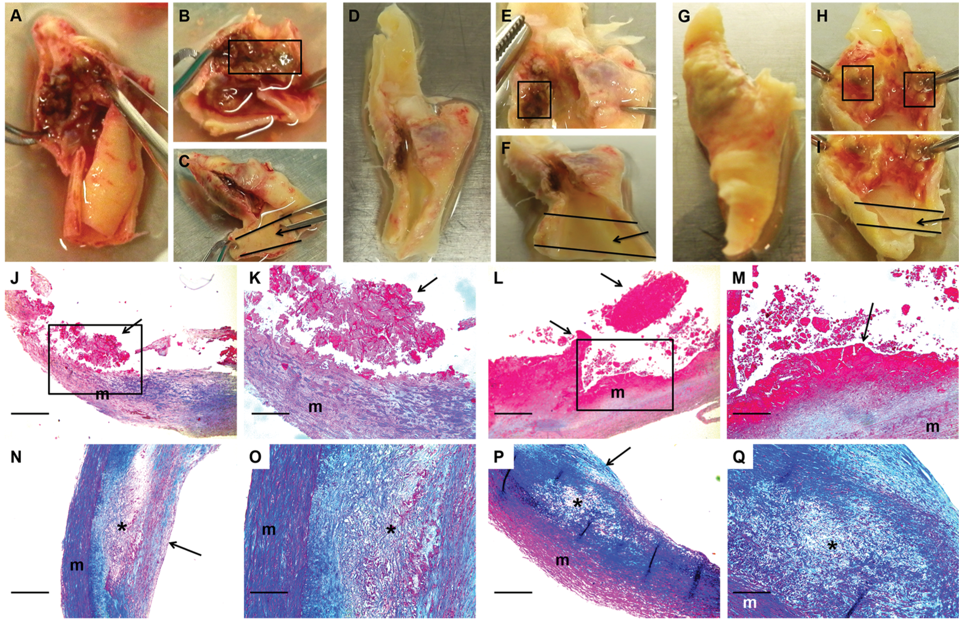
Figure 3. Macroscopic and histologic images of ruptured and stable segments of human carotid plaques.

Human carotid plaques were removed for clinical indications. Images are of 3 freshly harvested plaques (A-C, D-F, and G-I) and stained sections of 2 ruptured (J-K and L-M) and 2 stable (N-O and P-Q) plaques. Ruptured segments (boxes in B, E, and H) and stable segments (arrows in C, F, and I) were dissected free. A scalpel was used to cut thin slices from the caudal and cranial edges of the dissected segments. These slices were embedded in paraffin, and the remainder of each segment was used for protein extraction. Sections of 2 ruptured plaque segments (J-M; boxes in J and L are expanded in K and M) show disrupted intima with adherent thrombus (arrows; thrombus fragmentation is likely sectioning artifact). Sections of 2 stable plaque segments (N-O and P-Q; O and Q are expanded from N and P, respectively) show intact fibrous caps (arrows in N and P) and lipid-rich necrotic cores (asterisks) containing cholesterol clefts and foam cells. J-Q, m = vascular media. Size bars are 50 μm (J, L, N, and P) and 20 μm (K, M, O, and Q). J-Q, Masson’s trichrome stain.
