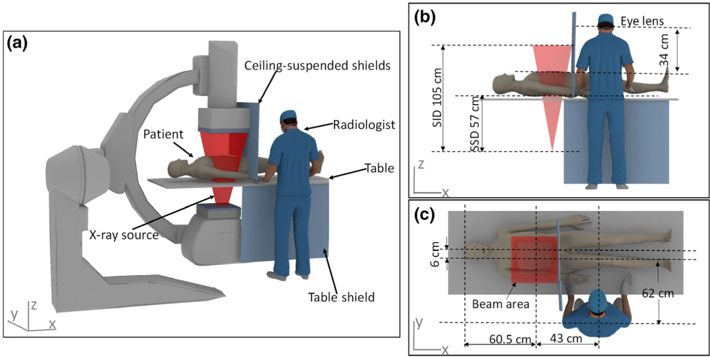
Fig. 1.

Geometric information for the simulation. (a) The interventional radiology suite consisting of two phantoms; (b) The side view showing the geometry of the x-ray source and the height of the eye lens relative to the top surface of the patient; (c) The top view showing the location of the radiologist and patient relative to the beam area.
