Fig. 3.
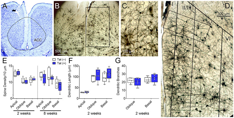
HIV-1 Tat exposure does not affect ACC layer V pyramidal neuron dendritic complexity or spine density. Dendritic morphology of ACC layer V pyramidal neurons of Tat(+) and Tat(−) mice were assessed after 2 or 8 weeks of DOX. Representative image of Nissl staining of the ACC (A). Representative images of Golgi-impregnated neurons in the ACC illustrating cortical layering (B&D) and layer V pyramidal neurons (C). Layer V pyramidal neurons (C) from Tat(−) and Tat(+) (not shown) mice displayed similar morphology and showed no differences in dendritic spine density (E) or dendritic complexity (F–G). Data are presented as mean ± SEM, multiple neurons (≥ 6) were sampled in each n = 5–7 mice per group; dashed-line, approximate borders of the ACC.

