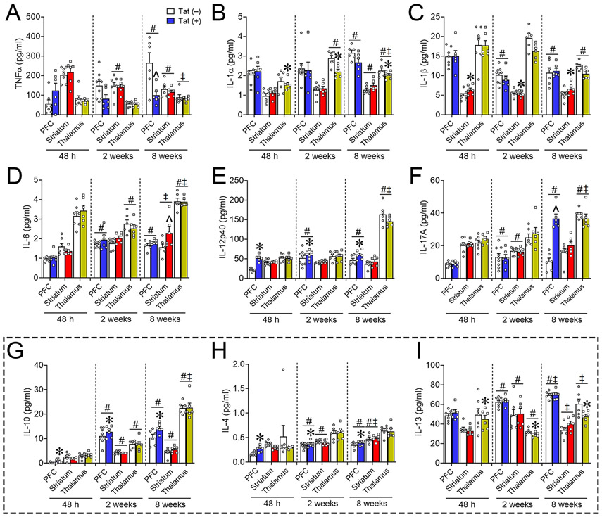
Fig. 8.
Proinflammatory and anti-inflammatory cytokine expression in the frontal cortico-BG-thalamocortical circuit is partially altered by HIV-1 Tat exposure. Proinflammatory and anti-inflammatory cytokine expression of Tat(+) (color bars) compared to Tat(−) (white bars) mice in the PFC, striatum, and thalamus after 48 h, 2 weeks, or 8 weeks of DOX exposure was largely unchanged. Proinflammatory IL-1β levels (C) were increased in the striatum after 48 h to 8 weeks of Tat exposure and IL-12p40 levels (E) were increased in the PFC; whereas in the thalamus IL-1α (B) levels were decreased. PFC expression of proinflammatory TNF-α (A) decreased, while IL-17A (F) increased in the PFC, and striatal IL-6 (D) expression increased in Tat(+) compared to Tat(−) mice after only 8 weeks of DOX. Expression of anti-inflammatory IL-10 (G) and IL-4 (H) was increased in the PFC; whereas IL-13 expression (I) was decreased in the thalamus after 48 h, 2 weeks, or 8 weeks of Tat exposure. Data are presented as mean ± SEM; n = 6–9 mice per group. Main effect, *p < 0.05 vs respective Tat(−) (control) within discrete regions. Main effect of exposure time within each separate region, #p < 0.05 vs respective 48 h exposure time; ‡p < 0.05 vs respective 2-week exposure time. ^p < 0.05, interaction of Tat and exposure time within discrete regions. Dashed line separates anti-inflammatory cytokines.

