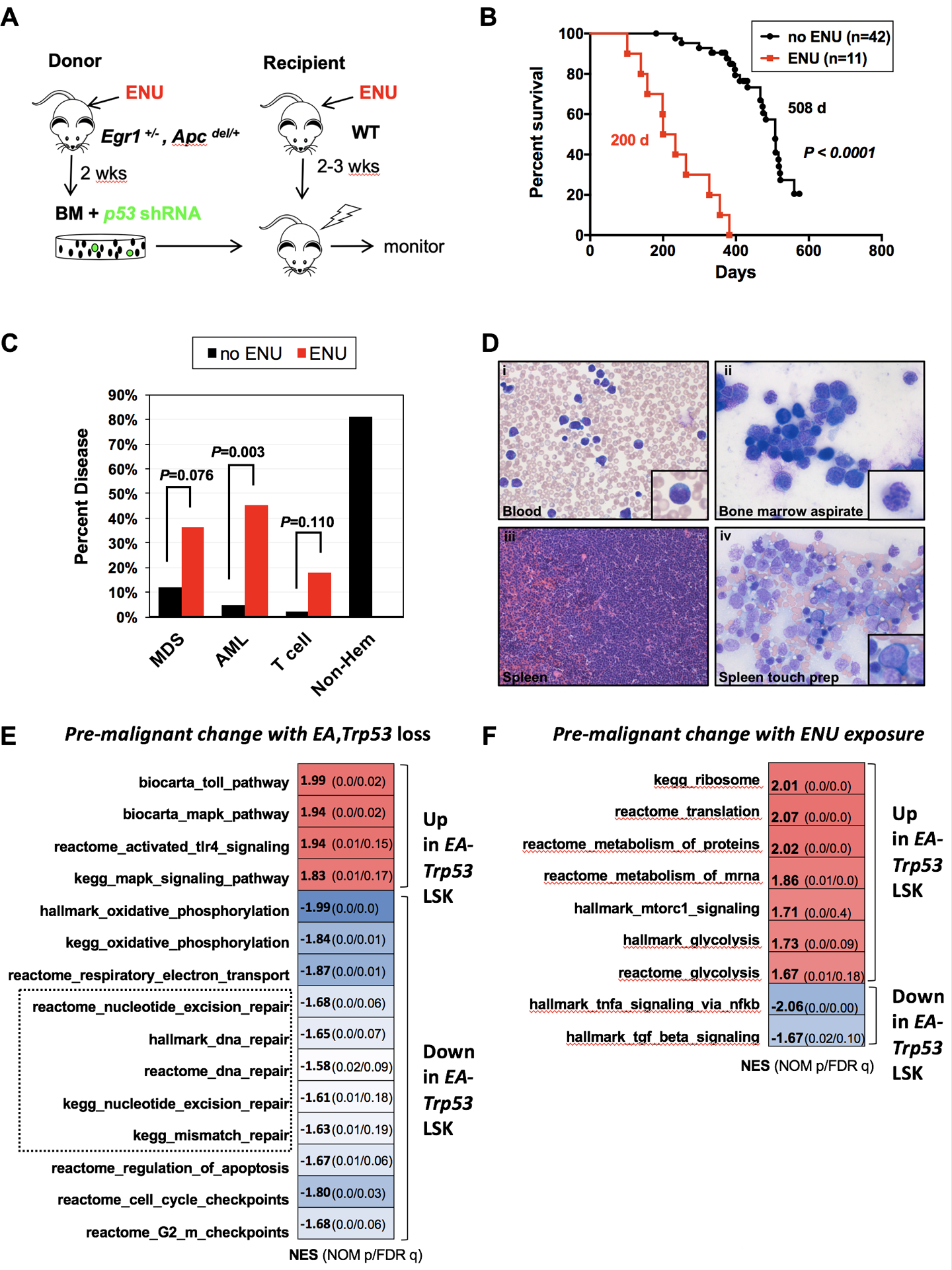
Fig. 1. Alkylating agent therapy increases the incidence of myeloid neoplasms.

(A) Schematic of ENU-treatment and transplantation scheme used to develop a mouse model of t-MN. Egr1+/−, Apcdel/+ bone marrow cells were transduced with Trp53 shRNA, and transplanted into lethally-irradiated WT recipient mice. For the ENU cohort, donor mice were treated once with 100 mg/kg ENU 2 weeks before bone marrow harvest; recipients were treated once with 100 mg/kg ENU, 2–3 weeks before lethal irradiation and transplantation. (B) Kaplan-Meier survival curves of untreated and ENU-treated mice. Percentage survival (time to sacrifice) is plotted vs. time in days. In the absence of ENU treatment, the mice survive significantly longer (508 d vs. 200 d, P <0.001), with a small percentage developing MDS/AML, but the majority succumb to advanced age and non-hematological effects. (C) Histological classification of diseases that arose in the no-ENU and ENU-treated mice. There is a significantly increased frequency of AML in the ENU-treated group. Most of the no-ENU mice died due to age-related issues, rather than hematopoietic malignancies. Disease frequency was compared using Fisher exact test. (D) Images of the myeloid disease in ENU-treated mice were obtained using an Olympus BX41 microscope (Melville, NY) and a 50x/0.9 (oil) or 40x/0.9 objective, and processed with Adobe Photoshop (San Jose, CA). Peripheral blood smears, bone marrow smears, and spleen touch preparations were stained with Wright–Giemsa (500x magnification), and spleen sections were stained with H&E (200x magnification). Examples of a myeloblast (inset-i), dysplastic granulocyte (inset-ii), sheets of infiltrating blasts (iii), and a dysplastic erythroid precursor (inset-iv) are shown. (E, F) LSK+ (Lin-, Sca1+, Kit+) cells were sorted from 3 different mouse cohorts: recipients of WT, luc shRNA+ bone marrow (controls) (n=3), recipients of Egr1+/−, Apcdel/+, Trp53 shRNA+ cells (EA-Trp53), either untreated (n=3) or treated with ENU (n=3), ~ 70–90 d after transplant, prior to the onset of overt leukemia. (E). GSEA of WT control (n=3) compared to EA-Trp53 LSK+ samples (includes both the no-ENU and ENU-treated groups (n=6)) to identify pre-malignant changes as a consequence of Egr1, Apc, and Trp53 loss. (F) GSEA of EA-Trp53 LSK+ samples from the no-ENU (n=3) versus ENU-treated (n=3) group was used to identify genetic consequences of in vivo ENU exposure. Biological pathways/processes that were significantly enriched (FDR<20%, nominal p value <0.05) in two or more Molecular Signatures Database (MSigDB) gene sets are shown. Heat map of the normalized enrichment scores (NES) shows that DDR pathways (DNA repair, apoptosis, checkpoints) are downregulated due to loss of Egr1, Apc, and Trp53, with or without ENU treatment. (E), and energy production pathways, such as mTORC1, protein translation and glycolysis, are upregulated as a consequence of ENU exposure (F).
