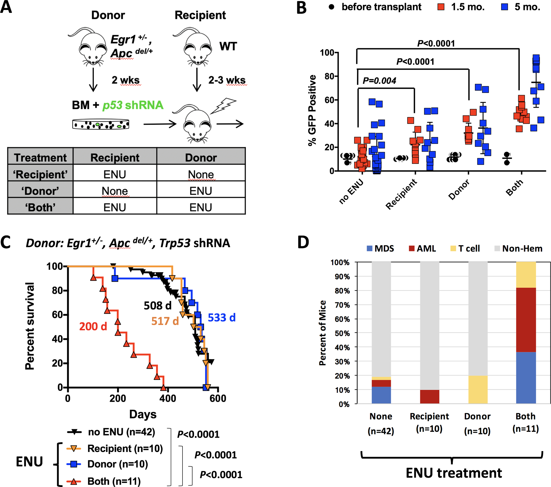
Fig. 2. Exposure of the bone marrow microenvironment to ENU plays a critical role in the development of MDS and AML.

(A) Schematic of the transplantation schemes used to elucidate the effects of alkylating agents on HSPCs, with Trp53 knockdown (donors), and bone marrow microenvironment (WT-recipients). Donor mice received one injection of ENU (100 mg/kg) two weeks before bone marrow harvest. Recipient mice received one injection of ENU (100 mg/kg) 3 weeks before lethal irradiation and transplantion. (B) Percentage of GFP+ cells in the bone marrow at the time of transplantation, and in the blood of mice at 1.5 and 5 months post-transplantation. There was a significantly greater percentage of GFP+ cells in the blood of ENU-treated mice at 1.5 month and 5 months. The P-values from a two-tailed Student t test at 1.5 months are shown. At 5 months, only ‘donor’ and ‘both’ conditions showed a significant expansion of GFP+ cells (P<0.0001). ENU exposure of both donor and recipient creates a more optimal environment for the expansion of Trp53 shRNA, GFP+ cells. (C) Kaplan-Meier survival curves of WT recipients transplanted with Egr1+/−, Apcdel/+ bone marrow cells transduced with Trp53 shRNA. When both donor and recipient mice are exposed to ENU, survival time is significantly decreased (200 d). There was no statistical difference in survival between ENU-donor vs. no ENU (P=0.831) or ENU-recipient vs. no ENU (P=0.737), or ENU-donor vs. ENU-recipient cohorts (P=0.910). (D) Histologic classification of diseases arising in the mice.
