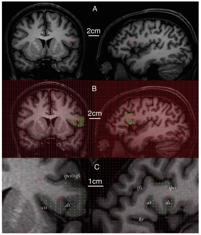
Figure 4.

Volume estimation of the frontal operculum in a human brain using stereology. The same voxel within the operculum is indicated by the red crosshair on each row. Only coronal (left) and sagittal (right) sections are used for illustrative purposes. A, Whole MR image without grid projection. B, Whole MR image with grid projection. Yellow points indicate the frontal operculum. C, Zoomed MR image focusing on the operculum (indicated by yellow points). ar, Anterior ascending ramus of the Sylvian fissure; ds, diagonal sulcus; hr, anterior horizontal ramus of the Sylvian fissure; ifs, inferior frontal sulcus; ipcs, inferior precentral sulcus; ts, triangular sulcus.
