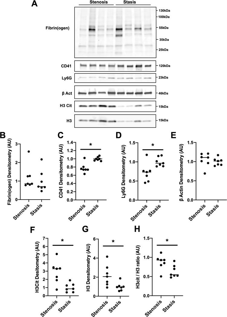
Figure 2: Composition of thrombi formed using the mouse IVC stenosis and stasis models of thrombosis.
Thrombus composition was assessed by western blotting of total soluble protein lysates from thrombi formed using the IVC stenosis and IVC stasis models of thrombosis (n=7 per group). (A) Representative western blots for fibrin(ogen), the platelet marker CD41, the neutrophil marker Ly6G, the cytoplasmic marker β Actin, citrullinated histone H3 (H3Cit) and the nuclear marker total histone H3 (H3) in thrombi formed using the IVC stenosis and IVC stasis models. Densitometric analysis of (B) fibrin(ogen), (C) CD41, (D) Ly6G, (E) β Actin, (F) H3Cit and (G) H3 in thrombi formed in the IVC stenosis model compared to the IVC stasis model. (H) The ratio of H3Cit to total H3 was significantly higher in thrombi formed in the IVC stenosis model compared to the IVC stasis model. * P<0.05, **P<0.01 Mann-Whitney U test. Data represented as individual values with a line for the median.

