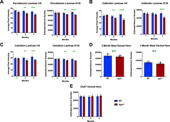
FIGURE 1 – Early interneuron loss in the spinal cords of Ppt1−/− mice.
(A-C) Analysis of lumbosacral cords immunostained for interneuron markers – parvalbumin, calbindin and calretinin respectively. Image densitometry (average pixel luminance) revealed a significant decrease in intensity of immunostaining for these markers in laminae I-III as early as 2 months for all antibodies stained, progressing at 3 months. Manual stereological counts of neurons positively immunostained for these markers in laminae IV-IX [98] revealed a significant loss of parvalbumin and calretinin positive interneurons as early as 2 months, and calbindin positive interneurons at 3 months. (D) Unbiased stereological estimates of neuron numbers from Nissl stained spinal cord sections showed no significant difference between WT (wildtype) and Ppt1−/− mice at 2 months (MO) in either the dorsal or ventral horns of the lumbosacral spinal cord. Two-tailed, unpaired, parametric t-test (n=5 mice/group). (E) Manual stereological estimates of Choline Acetyltransferase (ChAT) positive motor neurons showed no significant loss at any timepoint measured. P-values - **p≤0.01, ***p≤0.001, ****p≤0.0001; two-way ANOVA with post-hoc Bonferroni correction. Values shown are mean ± SEM. (n = 5 mice/group).

