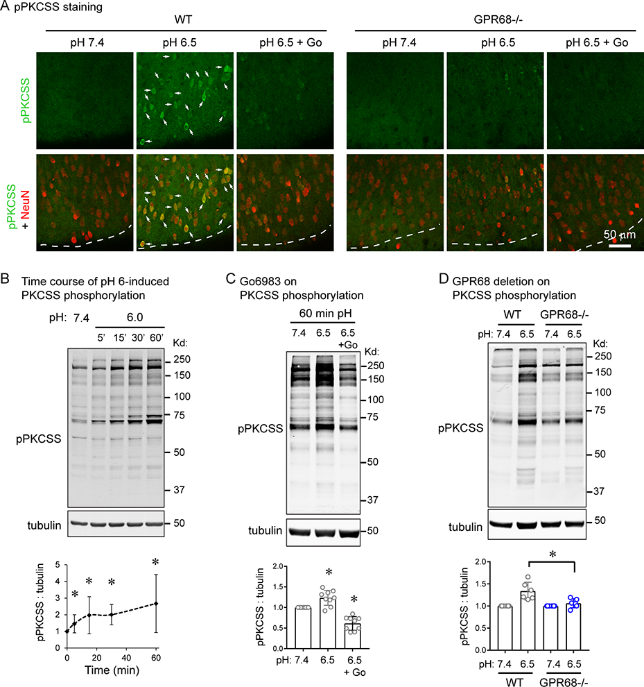
Figure 3. GPR68 mediates acidosis-induced signaling in slice neurons.
(A) Immunofluorescence of acidosis-induced phospho-PKCSS increase in cortical slices. WT and GPR68−/− slices were treated with pH 7.4, 6.5, or 6.5 + Go6983 for 15 min, and immunostained with a pPKCSS antibody together with a NeuN antibody. Arrows indicated neurons which exhibited an increase in pPKCSS signal in WT slices. Dashed lines indicate the outside (superficial) boundary of the cortex. (B) Representative Western blot and quantification of acidosis-induced PKC activation. Organotypic brain slices were treated with pH media as indicated, lysed, and analyzed by Western blot. To probe for PKC activation, a phospho-PKC substrate (pPKCSS) antibody was used. (C) Representative Western blot and quantification showing the effect of Go6983 on acid-induced phosphorylation of PKC substrate. (D) GPR68 deletion on acid-induced PKCSS phosphorylation. WT and GPR68−/− cortical slices were treated with pH media as indicated. Blots and images were representative from 4–7 different experiments.

