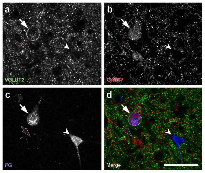
Fig. 6.
Identification of IC GAD67 neurons that project to the MGB. (a) One neuron with VGLUT2+ axosomatic endings (white arrow). (b) Large GAD67+ with axosomatic endings (white arrow) and a second GAD67+ neuron (black arrow) without axosomatic endings. (c) After a FG injection into the medial geniculate body, retrogradely labeled FG-positive cells were found in the IC (white arrows and arrowheads). (d) The large GAD67+ cells with VGLUT2+ axosomatic endings were labeled with FG (white arrow). GAD67+ neurons without VGLUT2+ axosomatic endings were not labeled with FG (black arrows). The majority of FG-positive cells are negative for GAD67 (white arrowheads). Green: VGLUT2; red: GAD67; blue: FG. Scale bar: 50 μm.

