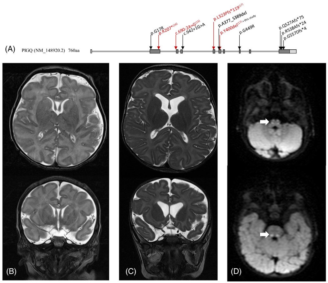
FIGURE 1.

A, Variants in PIGQ identified in this study localized on isoform 1 (NM_148920.2) of the gene. Variants reported in previous studies are in red with corresponding reference numbers in superscript. Brain MRI of subject 4 was performed at age 9 days, B, and 7 months, C. At each age, T2-weighted image sequences were performed with axial (top) and coronal (bottom) images shown. Brain MRI at 7 months showed progressive cortical volume loss with increased prominence of the lateral ventricles, consistent with loss of subcortical white matter volume compared to the prior study. Mild delay in myelination was apparent. D, Diffusion weighted axial images identified restricted diffusion in the medial lemniscus tracts, bilaterally
