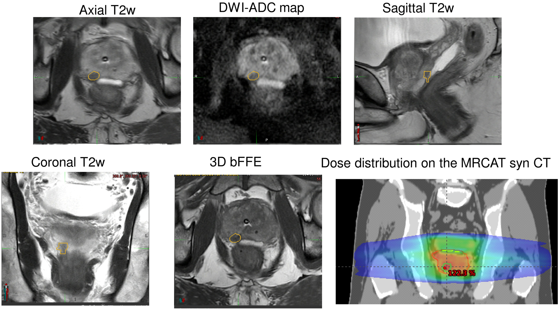
Figure 1:

MR-only workflow used to contour and plan dominant intraprostatic lesion (DIL). Anatomical (axial, coronal, sagittal T2 w MRI,3D bFFE goldseed) and apparent diffusion coefficient (ADC) maps derived from diffusion-weighted MRI are used to delineate dominate lesion. A Rx of 800×5 cGy with a boost of 900×5 cGy is planned.
