Figure 7.
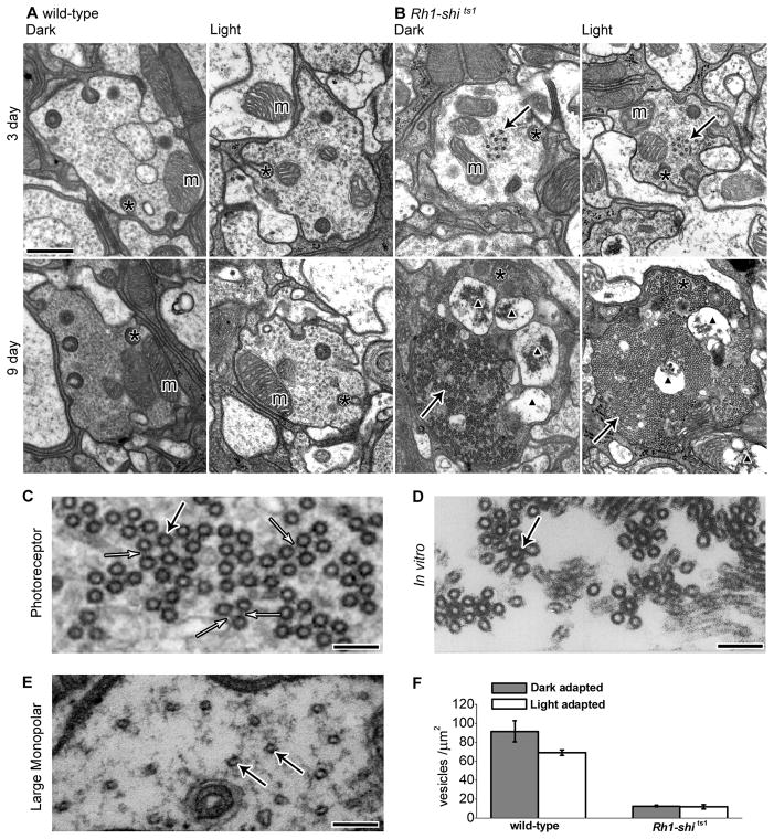
Overexpression of UAS-shits1 changes photoreceptor terminal morphology at 19°C. (A) EM cross sections of 3 and 9 days post-eclosion wild-type and (B) Rh1-shi ts1 terminals, reared at 18°C. Flies were exposed to 30 min of darkness (Dark) or to 30 min of darkness followed by 30 min of light (Light) at 18°C. Scale = 500 nm. Mitochondria (m), coated microtubules (solid arrows), capitate projections (asterisk), apoptotic mitochondria profiles (black triangle). (C) High magnification image of coated microtubules with cross-bridges (open arrow). (D) in vitro coated microtubules, reprinted from Cell, 59(3), Shpetner & Vallee, Identification of dynamin, a novel mechanochemical enzyme that mediates interactions between microtubules, 421–432, Copyright 1989, with permission from Elsevier and the authors. (E) Image of LMC wild-type microtubules. C–E Scale = 100 nm, microtubules (solid arrow). (F) Number of vesicles per μm2 for 3 days post-eclosion wild-type (dark- adapted; 91.40 ± 11.02, light-adapted; 68.86 ± 3.07) and Rh1-shi ts1 terminals (dark- adapted; 12.70 ± 0.78, light-adapted; 11.70 ± 2.26). Mean ± SEM given.

