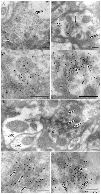
Figure 8.
Immunogold labeling confirms that the microtubule coat is dynamin. (A) Wild-type terminal showing 10 nm gold immuno-labeling in proximity to the membrane, indicated by open arrows and capitate projections (asterisk). (B) High magnification of A. (C) Photoreceptor terminal from Rh1-shi ts1, which in addition to labeling near capitate projections (open arrows), has labeling in the cytoplasm near coated microtubules (solid arrow). (D) High magnification of C. (E) Terminal from Rh1-shi ts1 showing labeling of oblique coated microtubules in the cytoplasm. Note that adjacent LMC profiles do not have cytoplasmic labeling, thus confirming the specificity of gold labeling to overexpressed shits1 dynamin. A–E Scale = 200 nm. (F) High magnification of labeled oblique and (G) cross section coated microtubules (solid arrow) showing that labeling extends to filaments adjacent (open arrow). F–G Scale = 100 nm.

